Dr. S. Sathish Babu
MBBS, MD . Professor & HOD
Overview
The Department of Radiodiagnosis, commonly known as the Radiology Department, plays a pivotal role in modern healthcare by utilizing advanced imaging techniques to diagnose, monitor, and guide the treatment of a wide range of medical conditions. The department’s primary objective is to provide accurate internal images of the body, aiding clinicians in precise diagnosis, effective treatment planning, and continuous monitoring of disease progression. Radiodiagnosis is integral to nearly all medical specialties, serving as a cornerstone for both preventive and curative healthcare practices, and enabling advanced treatment procedures with accuracy and confidence. The department is equipped with modern imaging modalities operated by skilled radiologists and technologists to ensure high-quality results. It emphasizes patient safety, timely reporting, and adherence to established imaging protocols. Through continuous technological upgrades and expertise, the department supports comprehensive, patient-centered clinical care.

Overview
The Department of Radiology at Sree Mookambika Institute of Medical Sciences (SMIMS) provides comprehensive diagnostic and interventional imaging services to support accurate diagnosis and effective patient management. Equipped with advanced imaging technology and a skilled team of radiologists and technicians, the department delivers high-quality imaging services across all clinical specialties. The department offers a wide range of diagnostic modalities including digital radiography, ultrasound, CT, MRI, mammography, and interventional procedures, ensuring timely and precise evaluation of medical conditions. Imaging services are integrated with the hospital’s PACS reporting system, enabling efficient image storage, reporting, and access to patient data. In addition to clinical services, the department plays a vital role in undergraduate and postgraduate medical education, research, and training, contributing to the advancement of diagnostic radiology and imaging sciences.

Dr. S. Sathish Babu
MBBS, MD . Professor & HOD
Overview
The Department of Radiodiagnosis, commonly known as the Radiology Department, plays a pivotal role in modern healthcare by utilizing advanced imaging techniques to diagnose, monitor, and guide the treatment of a wide range of medical conditions. The department’s primary objective is to provide accurate internal images of the body, aiding clinicians in precise diagnosis, effective treatment planning, and continuous monitoring of disease progression. Radiodiagnosis is integral to nearly all medical specialties, serving as a cornerstone for both preventive and curative healthcare practices, and enabling advanced treatment procedures with accuracy and confidence. The department is equipped with modern imaging modalities operated by skilled radiologists and technologists to ensure high-quality results. It emphasizes patient safety, timely reporting, and adherence to established imaging protocols. Through continuous technological upgrades and expertise, the department supports comprehensive, patient-centered clinical care.
Dr. S. Sathish Babu
MBBS, MD . Professor & HOD
Professor & HOD
Mission
To deliver accurate, timely, and safe diagnostic imaging services using advanced technology and expert interpretation to support precise clinical decision-making.
Contact Information
Email id:
radiology@sreemookambikainstitute.com
Faculties
| S.No | Name | Designation |
|---|---|---|
| 1 | Dr. S. Sathish Babu | Professor |
| 2 | Dr. Vinod Sukumaran | Professor |
| 3 | Dr. Rohit R Chennithala | Associate Professor |
| 4 | Dr. Bharath Chandran G | Assistant Professor |
| 5 | Dr. Najla P | Assistant Professor |
| 6 | Dr. O C Assvath | Assistant Professor |
| 7 | Dr. Jenish Radhakrishnan Sumathi | Assistant Professor |
| 8 | Dr. Vivek Kumar A S | Senior Resident |
| 9 | Dr. Jobin Mathew Jose | Senior Resident |
| 10 | Dr. Renees Thajudeen | Senior Resident |
| 11 | Dr. Siddharth Rupak S | Senior Resident |
| 12 | Dr. Linda Sherin G | Senior Resident |
| 13 | Dr. VISHNU SANKAR T S | Senior Resident |
| 14 | Dr. SUNILNITHI K | Junior Resident |
| 15 | Dr. Priyadharshini C | Junior Resident |
| 16 | Dr. Revanth | Junior Resident |
| 17 | Dr. Varshini V.M | Junior Resident |
| 18 | Dr. Roshan Shaji | Junior Resident |
| 19 | Dr. Avni S Kumar | Junior Resident |
| 20 | Dr. Sylviya Charles | Junior Resident |
| 21 | Dr. Dharshika Retna Sigamony | Junior Resident |
| 22 | Dr. Padmanaban | Junior Resident |
| 23 | Dr. Antlin Sushma Lysander | Junior Resident |
| 24 | Dr. Jevanish Nijin V H | Junior Resident |
| 25 | Dr. Dayaakar R | Junior Resident |
| 26 | Dr. Priyanka P S | Junior Resident |
| 27 | Dr. Rajashree.N.S | Junior Resident |
Infrastructure
1. SPECIALITY SERVICES
Imaging Services
X-ray: One of the most common imaging techniques, used for assessing bones, joints, chest, and other structures.
Computed Tomography (CT) Scan: Provides detailed cross-sectional images, especially useful for complex cases involving the brain, abdomen, and chest.
Magnetic Resonance Imaging (MRI): Uses strong magnetic fields and radio waves to generate detailed images, particularly for soft tissues such as the brain, spinal cord, and muscles.
Ultrasound (USG/Doppler): Uses sound waves to create images, commonly employed for monitoring pregnancies, abdominal organs, and blood flow studies.
Mammography: A specialized X-ray technique to examine the breasts and detect early signs of cancer.
Nuclear Medicine: Involves the use of small amounts of radioactive material to diagnose and treat diseases, often applied in cancer, bone disorders, and thyroid evaluations.
Speciality Services
Fluoroscopy: Continuous X-ray imaging that provides real-time visualization of internal structures, commonly used during interventional procedures such as catheter insertions and gastrointestinal studies
2.PROCEDURE
Interventional Radiology (IR): A subspecialty of radiology that performs minimally invasive procedures—such as biopsies, drainage, and stent placements—under imaging guidance using CT, ultrasound, or fluoroscopy.
3. POST GRADUATE PROGRAMMES/ FELLOWSHIPS
MD –Radiodiagnosis
4. CMEs, CONFERENCES, WORKSHOPS, SEMINARS DONE IN THE DEPARTMENT
Interdepartmental Meetings and Seminars
5. MERITS AND OTHER ACHIEVEMENTS
Strengths of the Department
Experienced Faculty: Highly qualified and experienced faculty members provide expert guidance and mentorship to students and researchers.
Diverse Research Areas: Specialization in multiple areas of Radiology, including Cardiac CT and Interventional Radiology, offering wide-ranging research opportunities.
Interdisciplinary Collaboration: Active collaboration with other departments fosters innovative research and knowledge exchange.
Educational Excellence: High-quality undergraduate and postgraduate programs prepare students for successful careers in academia, industry, and healthcare.
Achievements
Publication of Research Papers: Faculty and students publish in prestigious scientific journals, contributing to advancements in Radiology.
Awards and Recognition: Postgraduate students receive awards and recognition for best paper presentations and research contributions in academic conferences.
Community Outreach: Faculty and postgraduates participate in medical camps, educating the public and providing benefits through screening X-rays, mammograms, and ultrasounds.
Training and Mentorship: Ongoing training and mentorship on CBME (Competency-Based Medical Education) and research, nurturing the next generation of medical professionals.
Advanced Imaging Equipment
The Department of Radiology is equipped with modern diagnostic imaging systems to support comprehensive patient evaluation.
X-Ray Systems:
- 7 Static X-ray machines with varying capacities (300 mA – 800 mA)
- Digital Radiography (DR) system
- Computed Radiography (CR) system
- 7 Portable X-ray machines for bedside imaging
- Fluoroscopy imaging system for contrast studies
These systems support routine radiography, emergency imaging, and specialized contrast studies.
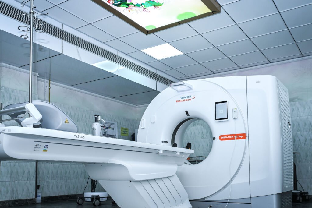
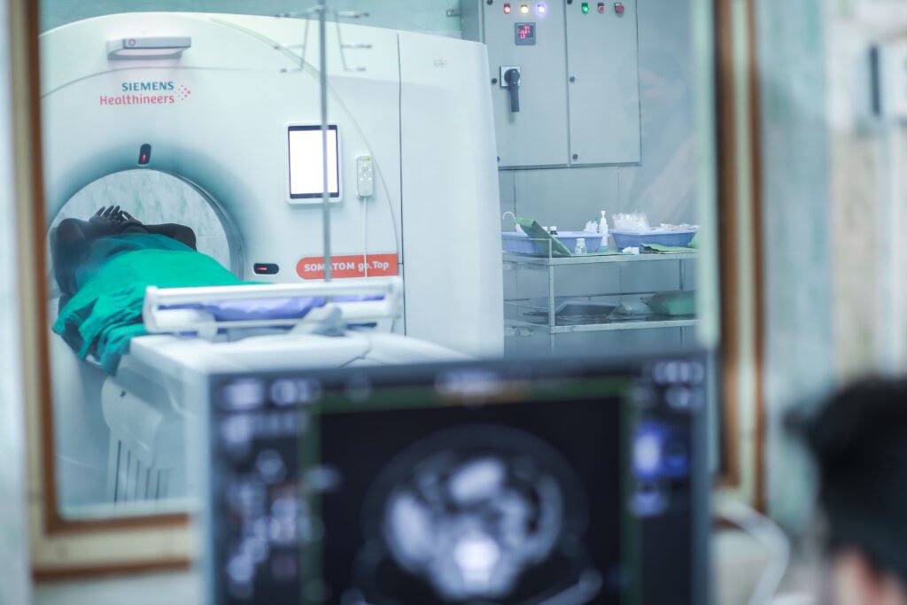
Computed Tomography (CT)
The department has two advanced CT scanners:
- 384-slice CT scanner (Siemens SOMATOM Go.Top) – advanced cardiac and neuro imaging capabilities
- 32-slice CT scanner (Siemens SOMATOM Scope)
These systems enable high-resolution imaging for trauma, oncology, vascular imaging, and multisystem evaluation.
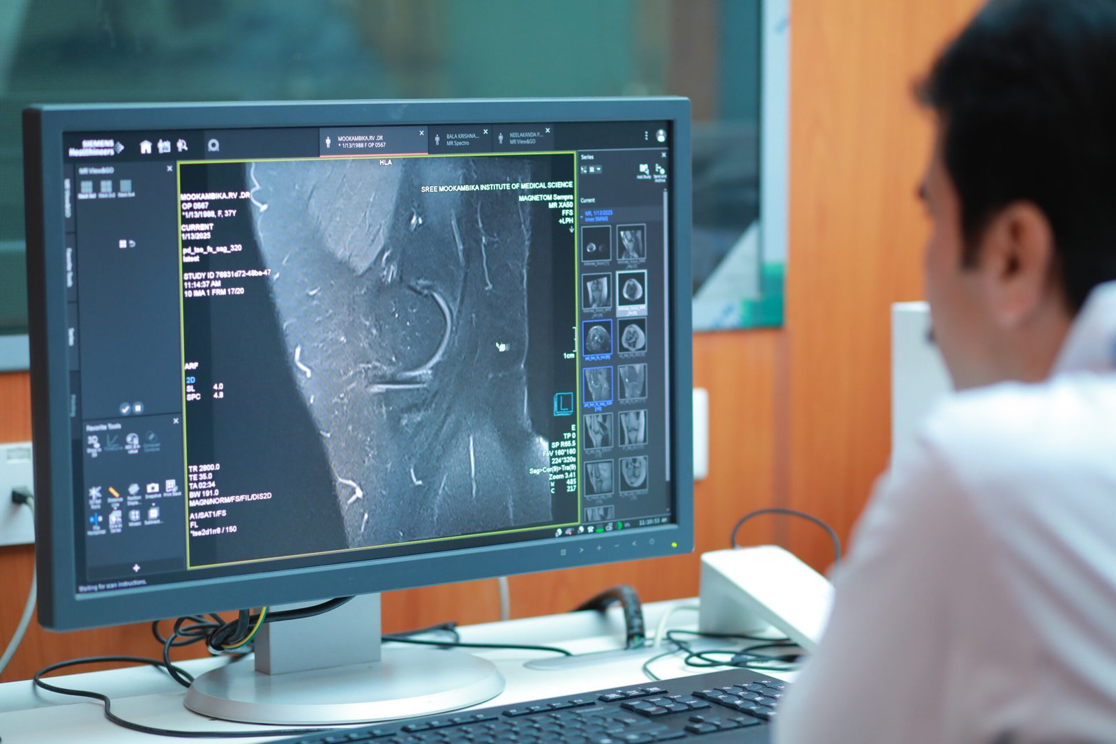
Magnetic Resonance Imaging (MRI)
- 1.5 Tesla MRI scanner – Siemens MAGNETOM Sempra
The MRI system supports detailed evaluation of neurological, musculoskeletal, abdominal, pelvic, and vascular conditions.
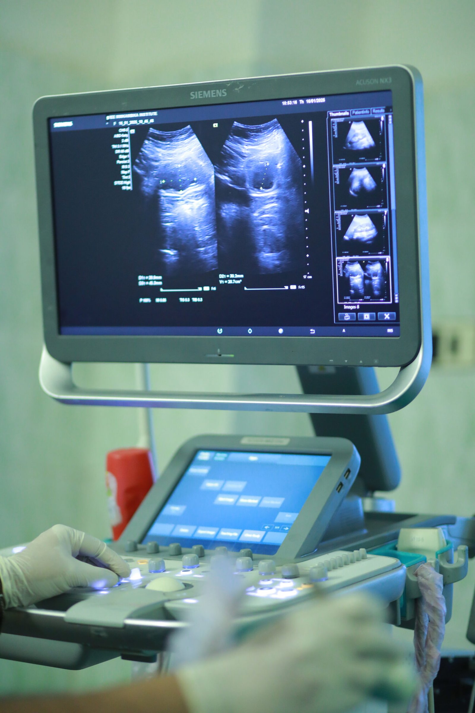
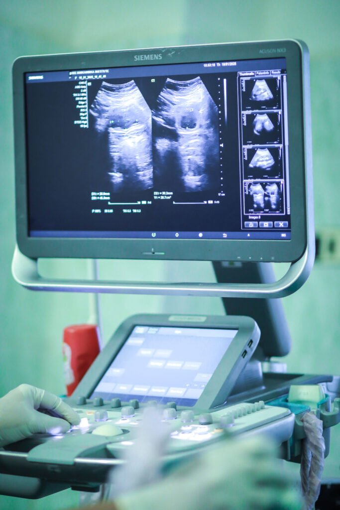
Ultrasound Systems
The department operates multiple advanced ultrasound machines including:
- Philips EPIC system with convex and 4D probe
- Siemens ACUSON NX3 with convex, linear, and phased probes
- Siemens ACUSON X300 systems for general imaging
These systems provide high-quality imaging for abdominal, obstetric, gynecologic, cardiac, and vascular examinations.
Doppler Imaging
Color Doppler ultrasound is used for evaluation of:
- Abdominal vascular conditions
- Peripheral vascular disease
- Carotid artery imaging
- Obstetric and fetal circulation studies
Specialized Imaging Systems
- Digital Mammography system – Siemens Mammomat
- Digital Subtraction Angiography (DSA) system – Siemens Artis Zee
- PET-CT scanner – Siemens Biograph Horizon
- Gamma Camera – Siemens Symbia EVO
- C-Arm systems for intraoperative imaging
Digital Imaging Infrastructure
- PACS Reporting System (MEDSYNAPSE)
- Integrated hospital information system for image storage and reporting
- Real-time access to radiology reports and images across the hospital network
Diagnostic and Interventional Services
The department provides a wide range of diagnostic and image-guided procedures including:
- Routine and specialized X-ray studies
- Contrast studies (IVP, barium swallow, barium meal, barium enema)
- Hysterosalpingography (HSG)
- Micturating cystourethrogram (MCUG)
- Ultrasonography and Doppler imaging
- USG-guided FNAC and biopsies
- Image-guided aspiration and drainage procedures
Advanced CT and MRI Studies
With advanced imaging technology and experienced radiologists, the Department of Radiology at SMIMS performs a wide spectrum of CT and MRI studies covering all major organ systems. These imaging modalities provide detailed anatomical and functional evaluation, supporting accurate diagnosis and clinical decision-making across multiple specialties.
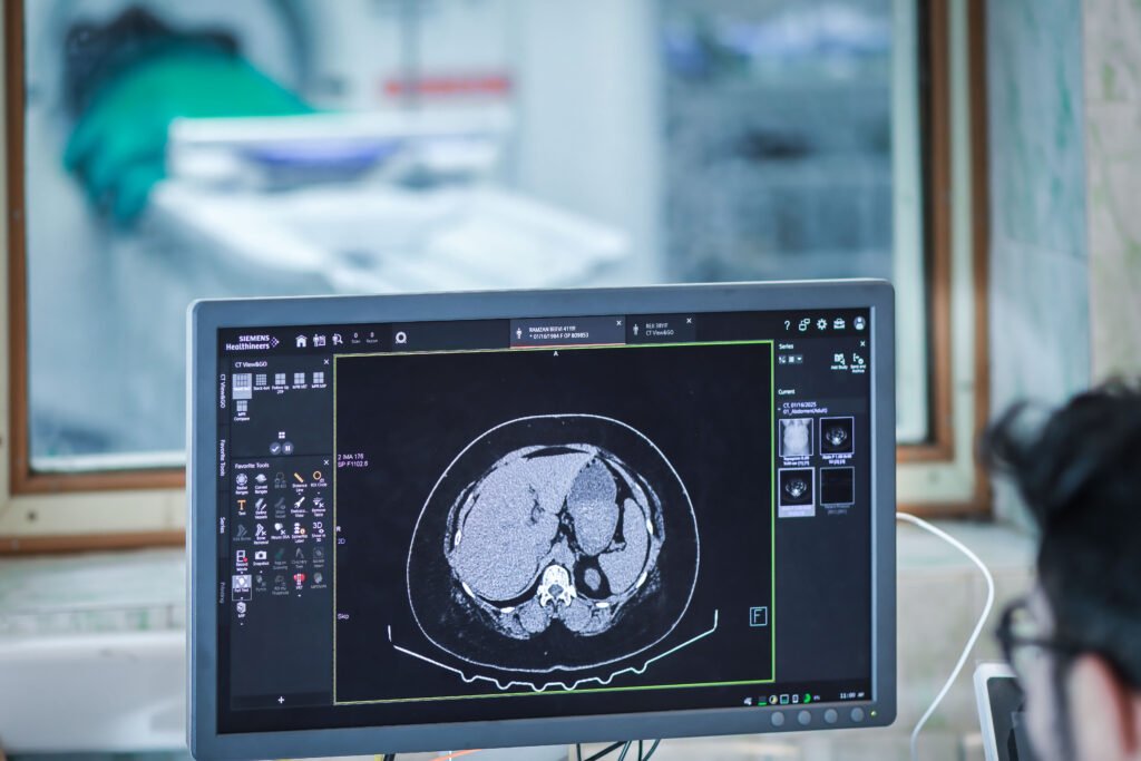
Computed Tomography (CT) Studies
The CT scanners enable high-resolution imaging for evaluation of trauma, oncology, vascular diseases, and multisystem conditions. The scope of CT imaging includes:
- CT Brain and Neuroimaging for stroke, trauma, tumors, and intracranial infections
- CT Chest and Lung Imaging for pulmonary infections, interstitial lung disease, and thoracic tumors
- CT Abdomen and Pelvis for hepatobiliary, gastrointestinal, pancreatic, renal, and urological conditions
- CT Enterography and CT Urography for gastrointestinal and urinary tract evaluation
- CT Angiography including coronary, carotid, cerebral, renal, pulmonary, and peripheral angiography
- CT-guided procedures including FNAC, biopsies, and drainage procedures
- CT for trauma evaluation and polytrauma assessment
Magnetic Resonance Imaging (MRI) Studies
The 1.5 Tesla MRI system provides detailed soft tissue imaging for neurological, musculoskeletal, abdominal, and vascular conditions. The scope of MRI includes:
- MRI Brain and Spine for neurological disorders, tumors, trauma, and degenerative conditions
- MRI for Stroke and Neurovascular evaluation
- MRI Abdomen and Pelvis for hepatobiliary, pancreatic, renal, and gynecological conditions
- MRI Musculoskeletal imaging for joints, ligaments, and soft tissue injuries
- MR Urography for evaluation of urinary tract disorders
- MR Cholangiopancreatography (MRCP) for biliary and pancreatic duct evaluation
- MRI for oncology imaging and tumor staging
These advanced imaging studies enable comprehensive diagnostic evaluation, early disease detection, and precise treatment planning for patients across multiple medical specialties.
Interventional Radiology Services
The Department of Radiology provides a range of image-guided interventional procedures that enable minimally invasive diagnosis and treatment of various medical conditions. These procedures are performed using advanced imaging modalities such as ultrasound, CT, and fluoroscopy, ensuring precision and patient safety.
Common interventional procedures include:
- Ultrasound-guided FNAC and biopsy
- CT-guided biopsy and aspiration procedures
- Image-guided drainage of abscesses and fluid collections
- Percutaneous catheter placements
- Diagnostic and therapeutic vascular procedures using Digital Subtraction Angiography (DSA)
- Renal Biopsies
- USG Guided IV/Arterial Cannulations
- Image-guided procedures for oncology diagnosis and management
These minimally invasive techniques reduce the need for open surgery, resulting in faster recovery, reduced complications, and improved patient outcomes.
Emergency and Trauma Imaging Services (24×7)
The Department of Radiology provides round-the-clock imaging services to support emergency and trauma care at SMIMS.
Emergency imaging services include:
- 24×7 X-ray services for trauma and emergency cases
- Emergency CT scans for head injury, stroke, polytrauma, and internal bleeding
- Ultrasound and FAST scans for trauma assessment
- Rapid imaging support for cardiac, neurological, and surgical emergencies
- Immediate radiology reporting integrated with the hospital’s PACS system
These services play a critical role in early diagnosis and timely management of life-threatening conditions, supporting the hospital’s emergency and critical care teams
Regulatory Compliance – AERB & PCPNDT
The Department of Radiology at Sree Mookambika Institute of Medical Sciences (SMIMS) strictly follows all national regulatory guidelines to ensure safe, ethical, and responsible use of diagnostic imaging services.
All radiological equipment and imaging facilities are operated in compliance with the regulations of the Atomic Energy Regulatory Board (AERB), Government of India, ensuring radiation safety for patients, healthcare workers, and the environment.
The department also strictly adheres to the provisions of the Pre-Conception and Pre-Natal Diagnostic Techniques (PCPNDT) Act, which regulates the use of ultrasound and other diagnostic techniques during pregnancy to prevent misuse for sex determination.
Key regulatory practices followed include:
- AERB-approved installation and operation of radiology equipment
- Regular radiation safety audits and quality assurance checks
- Personal dosimetry monitoring for radiology staff using TLD badges
- Use of protective radiation shielding devices such as lead aprons and barriers
- Appointment of a Radiation Safety Officer (RSO) to oversee radiation protection practices
- Strict compliance with PCPNDT registration and documentation requirements
- Maintenance of mandatory records and reporting protocols for obstetric ultrasound examinations
- Adherence to ethical guidelines ensuring no prenatal sex determination
Through strict compliance with AERB and PCPNDT regulations, the department ensures high standards of patient safety, ethical practice, and legal accountability in radiological services.
Radiation Safety
The Department of Radiology at Sree Mookambika Institute of Medical Sciences (SMIMS) maintains strict radiation protection practices to ensure the safety of patients, healthcare workers, and the environment. All imaging procedures involving radiation are conducted in accordance with national regulatory guidelines and internationally accepted radiation safety principles.
The department follows the ALARA (As Low As Reasonably Achievable) principle, ensuring that radiation exposure is minimized while maintaining optimal diagnostic image quality.
A designated Radiation Safety Officer (RSO) oversees all radiation safety measures, monitors compliance with safety regulations, and ensures that all equipment and procedures adhere to prescribed safety standards.
In addition, a Radiation Safety Committee functions within the institution to supervise radiation protection practices, review safety protocols, and ensure continuous monitoring of radiation exposure levels.
Key radiation safety measures include:
- Implementation of the ALARA principle to minimize radiation exposure
- Supervision and monitoring by a Radiation Safety Officer (RSO)
- Oversight by the Radiation Safety Committee
- Personal dosimetry monitoring (TLD badges) for staff working with radiation
- Use of protective shielding devices such as lead aprons, thyroid shields, and lead barriers
- Regular radiation safety training and awareness programs for staff
- Periodic equipment quality assurance and radiation audits
These measures ensure that all radiological procedures are performed with the highest standards of safety, responsibility, and regulatory compliance.
Teaching and Academic Activities
The Department of Radiology actively contributes to medical education and training.
- Postgraduate seats: 6 MD (Radio Diagnosis) per year
- Clinical training for MBBS students through demonstrations and imaging discussions
- Case discussions, seminars, and journal clubs
- Training in interpretation of advanced imaging modalities
- Participation in CME programs, conferences, and academic research
The department aims to train competent radiologists skilled in modern diagnostic imaging techniques and clinical decision support.
Regulatory Compliance – AERB & PCPNDT
The Department of Radiology at Sree Mookambika Institute of Medical Sciences (SMIMS) strictly follows all national regulatory guidelines to ensure safe, ethical, and responsible use of diagnostic imaging services.
All radiological equipment and imaging facilities are operated in compliance with the regulations of the Atomic Energy Regulatory Board (AERB), Government of India, ensuring radiation safety for patients, healthcare workers, and the environment.
The department also strictly adheres to the provisions of the Pre-Conception and Pre-Natal Diagnostic Techniques (PCPNDT) Act, which regulates the use of ultrasound and other diagnostic techniques during pregnancy to prevent misuse for sex determination.