
Oral Medicine & Radiology
Diagnosis and management of oral manifestations of systemic disease — empowered by CBCT, digital OPG and the full spectrum of maxillofacial imaging modalities.
Diagnose. Image. Manage.
The department deals with diagnosis and management of oral manifestations of systemic disease and the care of medically compromised patients. The faculty specialise in temporomandibular disorders and radiographic interpretation of complex oro-facial conditions.
Oral & Maxillofacial Radiology is a dental specialty concerned with the creation and interpretation of images using all modalities of radiant energy to diagnose diseases of the oro-facial region. Digital imaging, CT, MRI and CBCT are now part of the dentist's arsenal — partly due to the work of oral & maxillofacial radiologists.
An exclusive Cone Beam Computed Tomography centre, staffed by faculty qualified in CBCT, defines the new face of dental practice the world is warming up to.
What we set out to achieve
Equip dental researchers with modern facilities to meet evolving oral health care needs.
Promote laboratory and clinical research collaboration across key thematic areas.
Develop clinically effective treatment at both public-health and individual-patient levels.
Cross-cutting activity that improves both clinical practice and population oral health.
Our team
| Name | Designation |
|---|---|
| Dr. Suresh C.S, MDS | Professor & HOD |
| Dr. Lakshmi P.S, MDS | Reader |
| Dr. Dhanya S.V, MDS | Reader |
| Dr. Sweety S. Davis, MDS | Senior Lecturer |
| Dr. S. Jeflin Steni, MDS | Senior Lecturer |
Advanced imaging, end-to-end
Our department houses dedicated infrastructure for advanced imaging — including the CBCT machine and OPG machine. CBCT has shown a tremendous upward trend since the late 1990s and lets clinicians view and analyse osseous oral and maxillofacial structures in three dimensions, overcoming the magnification and superimposition issues of 2D modalities.
The Planmeca ProMax® 3D (made in Finland) is an all-in-one imaging family with up to five modalities — meeting every modern maxillofacial imaging need. Its unique Planmeca Ultra Low Dose™ protocol enables CBCT imaging with a lower effective patient dose than a standard 2D panoramic exposure.
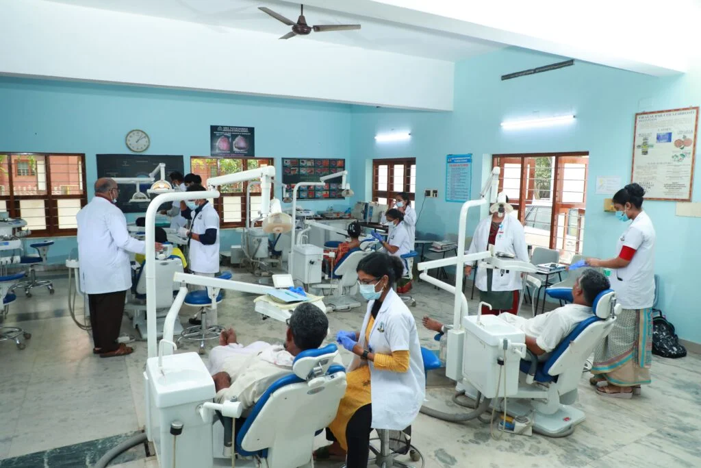
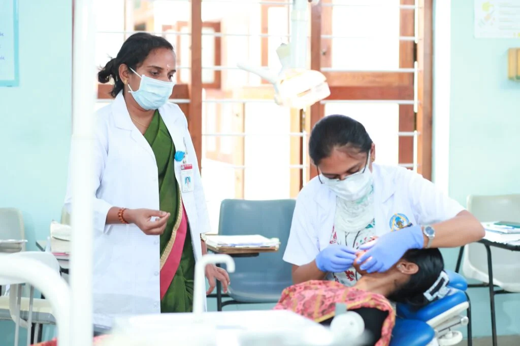
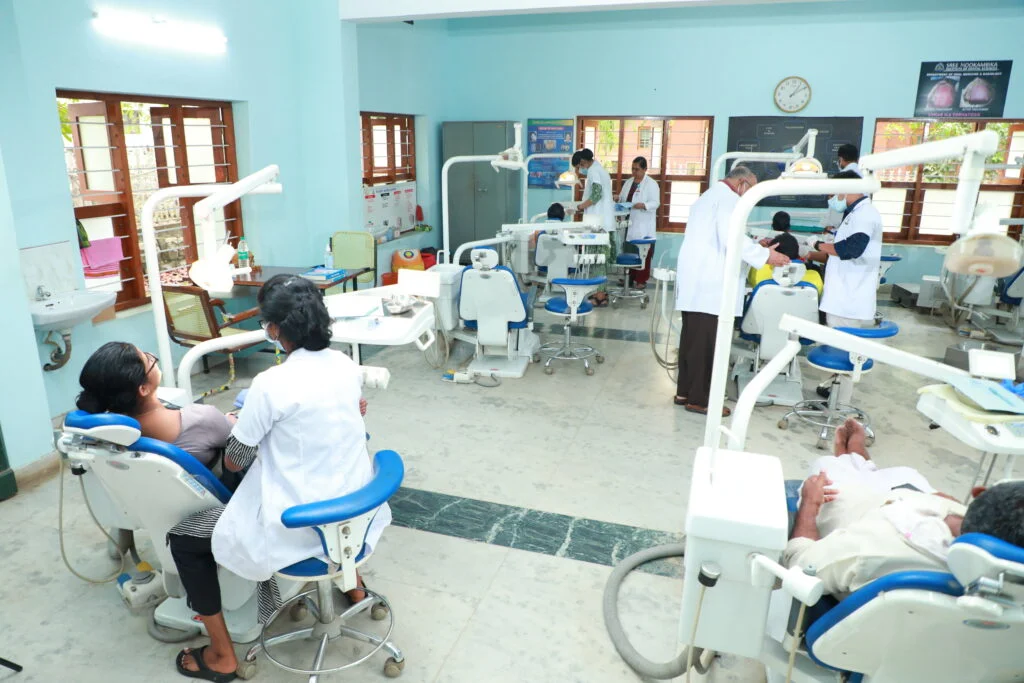
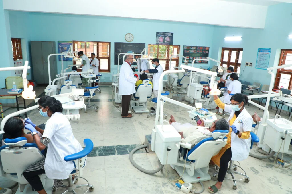
Inside the department
Overview
Methodology
Special lectures by eminent faculty from other universities on specific topics.
Problem-based learning to give students a 'real life' clinical experience.
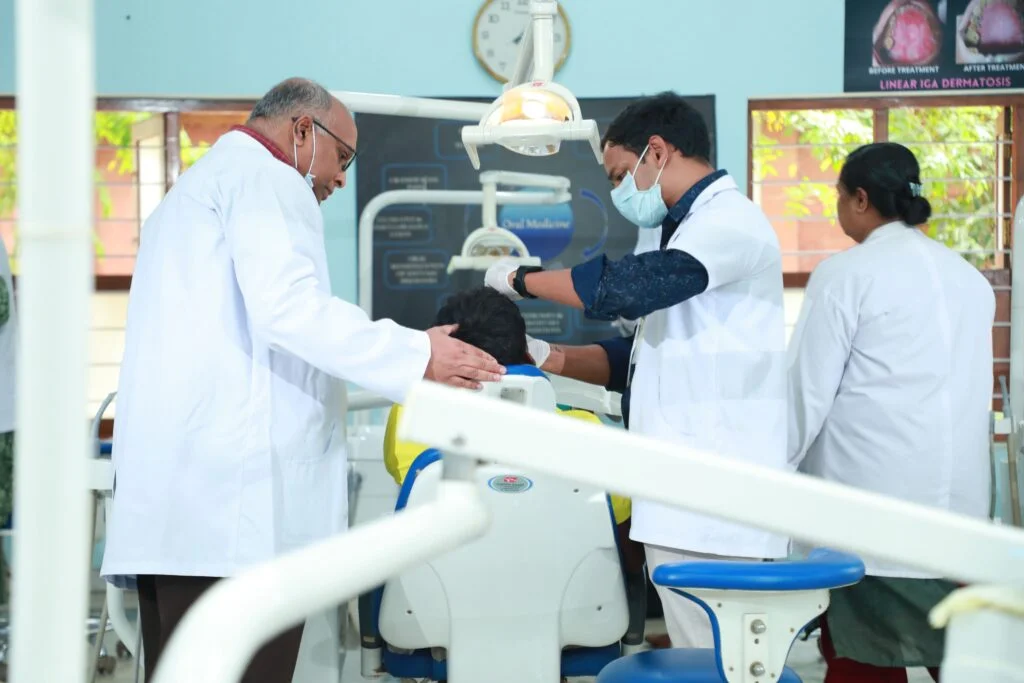
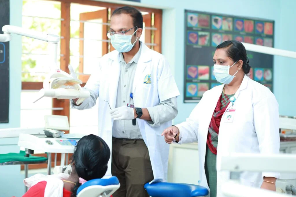
Chair-side teaching for a thorough hands-on clinical approach.
Open faculty accessibility — no student left shy or introverted.
Student-managed UG & PG notice boards evaluated for content and quality.
Periodic student and patient feedback shapes department direction.
Internal theory, oral and practical assessments for UG and PG.
Regular intra-department staff meetings tracking academic progress.
Diagnostic & therapeutic scope
Detection of oral cancer and precancerous lesions.
Identification of oral manifestations of systemic disorders.
Diagnosis and medical management of various oral diseases.
Diagnosis & non-surgical management of temporomandibular disorders.
Medical management of complex orofacial pain conditions.
Diagnosis & medical management of salivary gland diseases.
Comprehensive management of medically compromised patients.
Minor biopsy procedures for definitive diagnosis.
Equipment & clinical services
Cone Beam Computed Tomography for 3D maxillofacial imaging.
100 kVp digital extra-oral radiographic machine.
Panoramic and cephalometric digital imaging.
Photo-stimulable phosphor plate imaging.
High-resolution intra-oral radiography.
Real-time chairside visual diagnosis.
Imaging of salivary gland ducts and pathology.
Retrieval of salivary calculi (lith retrieval).
Tissue staining for early lesion detection.
Dedicated clinic for temporomandibular joint disorders.
Non-invasive detection of cracks and caries.
Multidisciplinary review of complex oncology cases.
CDEs, Achievements & Publications
Continuing Dental Education programmes, workshops and academic events conducted by the department.
Faculty and student awards, recognitions and milestones across national and international platforms.
Peer-reviewed publications and scientific contributions from the department.