
Foundation of clinical excellence.
The Department of Anatomy provides a strong foundation in the structural organization of the human body through gross anatomy, histology, embryology and genetics. Teaching blends regional, systemic, clinical, cross-sectional and radiological approaches so every student moves from structure to function to bedside relevance.
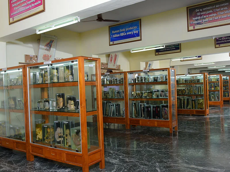
Students gain hands-on skills in cadaver dissection, preservation and histological techniques in a department equipped with a fully air-conditioned lecture hall, two demonstration rooms, dedicated histology and dissection halls, an embalming room, a 500+ specimen museum and a research laboratory with advanced microscopes.
Mission
To develop competent, ethical healthcare professionals with a strong foundation in anatomical sciences and lifelong learning.
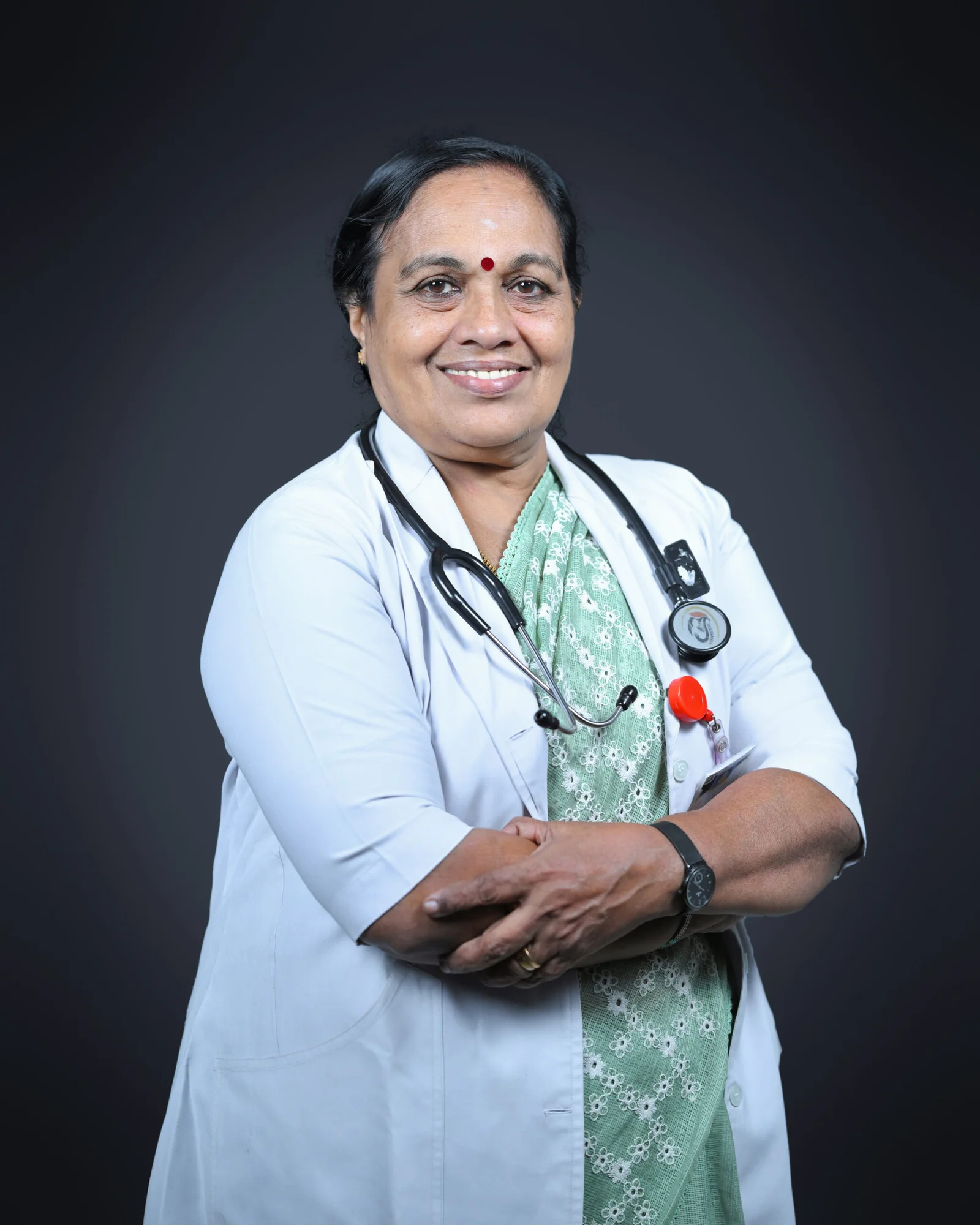
Leadership
Dr. Girija Kumari K
MBBS, MD — Professor & HOD
Faculty
A team of 16 educators shaping anatomical thinking.
| # | Name | Designation |
|---|---|---|
| 01 | Dr. Lathi Kumari K | Professor |
| 02 | Dr. Sneha Guruprasad Kalthur | Professor |
| 03 | Dr. Girija Kumari Kamalamma | Professor |
| 04 | Dr. Rathija Sree Kumar Prasanna Kumari | Associate Professor |
| 05 | Dr. Susie Jeyalyn David | Assistant Professor |
| 06 | Dr. Kalai Selvi C | Assistant Professor |
| 07 | Dr. Rincy Gomas | Senior Resident |
| 08 | Dr. Binija R | Tutor |
| 09 | Dr. Thishon Kumar T S | Tutor |
| 10 | Dr. Delima Antony | Tutor |
| 11 | Dr. Rathisha Sreekumar | Tutor |
| 12 | Dr. Jithu C T | Tutor |
| 13 | Dr. Jibin Hans V N | Tutor |
| 14 | Dr. Sivaduth Chandran | Tutor |
| 15 | Dr. Farhana A J | Tutor |
| 16 | Dr. Sujith J | Tutor |
Infrastructure
Spaces designed for discovery.
From the prosected dissection hall to a 500-specimen museum and a research lab with advanced microscopy, every facility is built around active, hands-on learning.
AC Lecture Hall
Fully air-conditioned with audiovisual aids and two demonstration rooms.
Histology Lab
Individual microscopes and slide projectors for every student.
Dissection Hall
Prosected specimens and skeleton models for hands-on learning.
Embalming Room
Dedicated preservation facility supporting cadaveric study.
Anatomy Museum
500+ wet specimens, charts, 3D models and rare congenital specimens.
Research Lab
Advanced microscopes supporting faculty and PG research.
Departmental Library
Textbooks, references and digital resources for self-study.
Cold Storage
Specialty cold-storage for cadaver preservation beyond mortuary.
Procedures
Embalming and histology section preparation are conducted in-house, training students in the foundational techniques of preservation and tissue study.
Postgraduate Programme
Academic activities are scheduled as per NMC norms — seminars, tutorials, discussions, journal clubs, peripheral postings and active PG research work.
CMEs & Academic Programs
Continuous learning, every year.
- Seminars, tutorials, group discussions and journal clubs as per NMC norms
- Peripheral postings and structured PG dissertation work
- Two CMEs conducted — Clinical Anatomy of Peripheral Nerves (2020) and Embryo ART (2023)
- World Anatomy Day observed annually (2022, 2023, 2024)
- Guest Lecture — Learning Anatomy: Blending Tradition with Technology (Oct 2024)
Student Activities
Where curiosity meets creativity.
Merits & Achievements
Faculty and PGs regularly participate in National and International conferences as chairpersons and speakers.