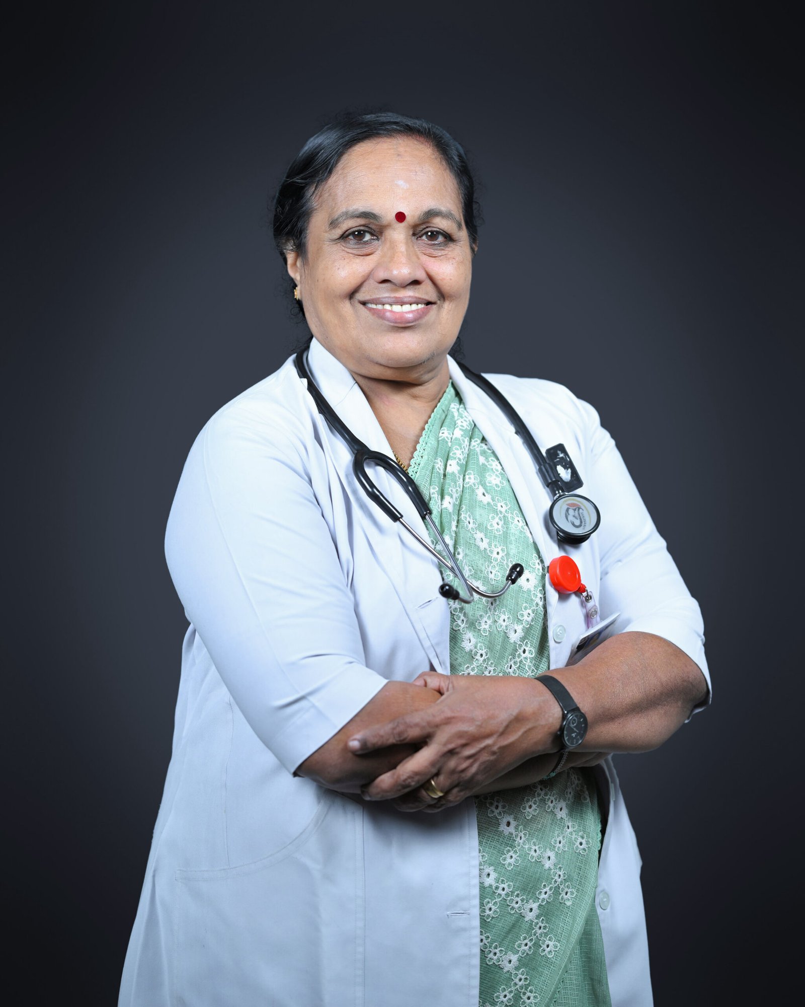
Dr. Girija Kumari. K
MBBS, MD
Professor & HOD
Overview
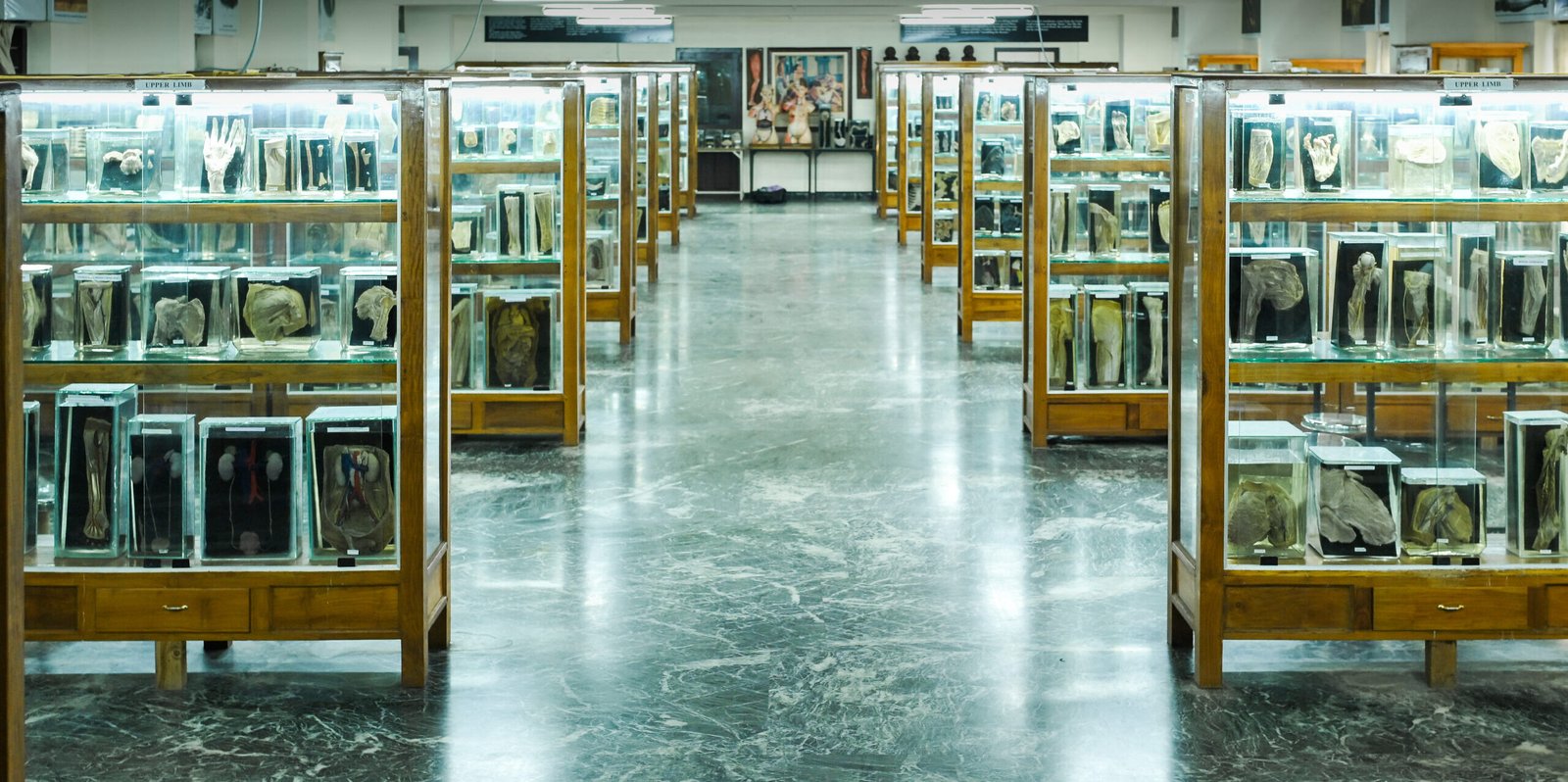
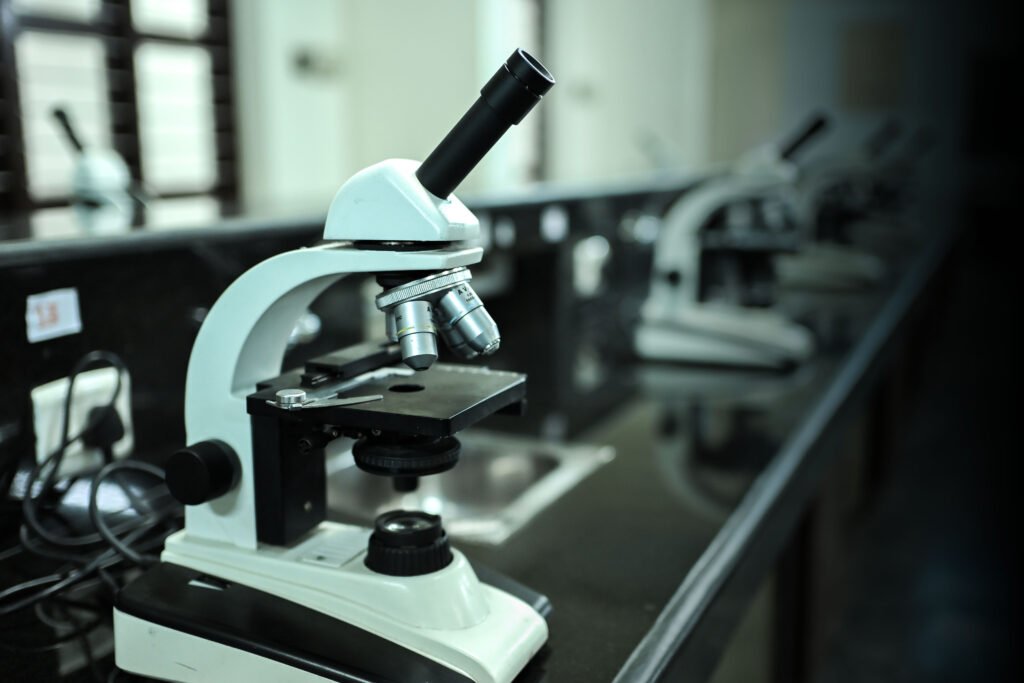
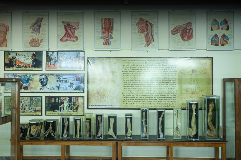
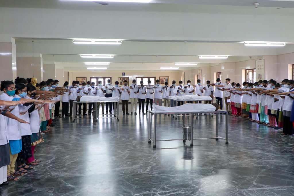
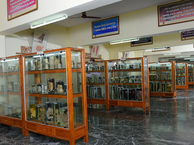
The Department of Anatomy provides a strong foundation in the structural organization of the human body through gross anatomy, histology, embryology, and genetics, taught using regional, systemic, clinical, cross-sectional, and radiological approaches. Students gain hands-on skills in cadaver dissection, preservation, and histological techniques. The department is equipped with a fully air-conditioned lecture hall with audiovisual aids, two demonstration rooms, a histology lab with individual microscopes and slide projectors, and a dissection hall containing prosected specimens and skeleton models. It also includes an embalming room, a museum with over 500 wet specimens, charts, 3D models, and rare congenital specimens, and a research laboratory with advanced microscopes. A well-organized departmental library with textbooks, reference materials, and digital resources supports effective self-study and research.
Overview
The Department of Anatomy provides a strong foundation in the structural organization of the human body through gross anatomy, histology, embryology, and genetics, taught using regional, systemic, clinical, cross-sectional, and radiological approaches. Students gain hands-on skills in cadaver dissection, preservation, and histological techniques. The department is equipped with a fully air-conditioned lecture hall with audiovisual aids, two demonstration rooms, a histology lab with individual microscopes and slide projectors, and a dissection hall containing prosected specimens and skeleton models. It also includes an embalming room, a museum with over 500 wet specimens, charts, 3D models, and rare congenital specimens, and a research laboratory with advanced microscopes. A well-organized departmental library with textbooks, reference materials, and digital resources supports effective self-study and research.
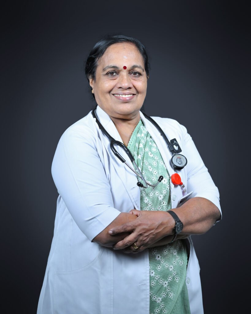
Dr. Girija Kumari. K ,
MBBS, MD
Professor & HOD
Overview
The Department of Anatomy provides a strong foundation in the structural organization of the human body through gross anatomy, histology, embryology, and genetics, taught using regional, systemic, clinical, cross-sectional, and radiological approaches. Students gain hands-on skills in cadaver dissection, preservation, and histological techniques. The department is equipped with a fully air-conditioned lecture hall with audiovisual aids, two demonstration rooms, a histology lab with individual microscopes and slide projectors, and a dissection hall containing prosected specimens and skeleton models. It also includes an embalming room, a museum with over 500 wet specimens, charts, 3D models, and rare congenital specimens, and a research laboratory with advanced microscopes. A well-organized departmental library with textbooks, reference materials, and digital resources supports effective self-study and research.
Dr. Girija Kumari. K
MBBS, MD . Professor & HOD
Professor & HOD
Mission
To develop competent, ethical healthcare professionals with a strong foundation in anatomical sciences and lifelong learning.
Contact Information
Email id:
anatomy@sreemookambikainstitute.com
Contact Information
Email id:
anatomy@sreemookambikainstitute.com
Faculties
| S.No | Name | Designation |
|---|---|---|
| 1 | Dr.Lathi Kumari K | Professor |
| 2 | Dr. Sneha Guruprasad Kalthur | Professor |
| 3 | Dr.Girija Kumari Kamalamma | Professor |
| 4 | Dr.Rathija Sree Kumar Prasanna Kumari | Associate Professor |
| 5 | Dr.Susie Jeyalyn David | Assistant Professor |
| 6 | Dr.Kalai Selvi C | Assistant Professor |
| 7 | Dr.RINCY GOMAS | Senior Resident |
| 8 | Dr.Binija R | Tutor |
| 9 | Dr.Thishon Kumar T S | Tutor |
| 10 | Dr.Delima Antony | Tutor |
| 11 | Dr.Rathisha Sreekumar | Tutor |
| 12 | Dr.Jithu C T | Tutor |
| 13 | Dr.Jibin Hans V N | Tutor |
| 14 | Dr.Sivaduth Chandran | Tutor |
| 15 | Dr.Farhana A J | Tutor |
| 16 | Dr.Sujith J | Tutor |
Introduction to Anatomy
Anatomy is a fundamental branch of medical science that focuses on understanding the structural organization of the human body. It includes various subdivisions such as gross anatomy, which deals with structures visible to the naked eye; microscopic anatomy or histology, which studies tissues; and developmental anatomy or embryology, which explains human growth and development. The subject also incorporates genetics and uses different approaches like regional, systemic, clinical, cross-sectional, and radiological anatomy. Practical components such as cadaver dissection and tissue preservation play a vital role in enhancing students’ understanding.
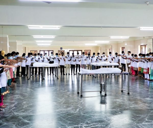
Objectives of Studying Anatomy
The primary aim of teaching anatomy is to provide students with a thorough knowledge of the structure of the human body at both gross and microscopic levels. It helps students understand the relationship between structure and function and recognize the clinical significance of anatomical features. By studying anatomy, students learn to identify body systems, understand how they function, and relate anatomical knowledge to disease conditions and medical practice.
Clinical Relevance and Integration
Anatomy education emphasizes clinical application by integrating theoretical knowledge with practical understanding. Students learn about the spatial relationships of body structures, functional anatomy, and cross-sectional views that are essential for diagnosis and treatment. The study of tissues is correlated with physiological functions and pathological changes. Special attention is given to the nervous system to understand how structural damage can affect body functions. Embryology helps explain congenital abnormalities and developmental variations.
Facilities and Infrastructure
The department is equipped with modern facilities to support effective learning. These include well-designed lecture halls with audiovisual aids, dissection halls for hands-on learning, and histology laboratories with microscopes and prepared slides. Museums displaying preserved specimens and models enhance visual learning. Additional facilities such as embalming rooms, research laboratories, and departmental libraries provide a comprehensive academic environment for students.
Teaching Methods and Student Support
Teaching methods are designed to promote active learning and better understanding. Techniques such as group discussions, seminars, case-based learning, and guest lectures by clinicians help bridge the gap between theory and practice. Horizontal integration with other subjects ensures a multidisciplinary approach. Regular internal assessments and examinations track student progress, while additional support is provided to slow learners through remedial classes and guidance.
Conclusion
The study of anatomy forms the foundation of medical education by combining theoretical knowledge, practical skills, and clinical relevance. It enables students to understand the human body in both normal and diseased conditions, preparing them for advanced medical studies and professional practice in healthcare.
1. SPECIALITY SERVICES
2. PROCEDURE
3. POST GRADUATE PROGRAMMES/ FELLOWSHIPS
4. CMEs, CONFERENCES, WORKSHOPS, SEMINARS DONE IN THE DEPARTMENT
Two CMEs conducted by Department
14/02/2020 – Recent Updates in the Clinical Anatomy of Peripheral Nerves
30/06/2023 – Embryo ART
World Anatomy Day observed – 15th October 2022, 2023, 2024
15/10/2024 – Guest Lecture: Learning Anatomy – Blending Tradition with Technology by Dr. Doris, GMC TVM
Student Activities
Model Making Competition – 2022
Quiz Competition – 2023 (Clinical Anatomy Knowledge for all phases)
Body Painting
Embryo Fest – Embryology Model Making Competition 2023
5. MERITS AND OTHER ACHIEVEMENTS