Overview
The Department deals with the diagnosis and management of oral manifestations of systemic disease and management of medically compromised patients. The faculty also specializes in management of Temporo-mandibular disorders and radiographic interpretation of complex Oro-facial disorders. Oral and Maxillofacial Radiology is a dental specialty and radiology discipline concerned with the creation and interpretation of images and data generated by all modalities of radiant energy used to diagnose and manage diseases, disorders, and conditions of the oral and maxillofacial region. Recent advancements in imaging science have enabled dentists to give far better diagnostic services to their patients while exposing them to the least amount of radiation possible, using modalities that were previously unknown. Digital imaging, CT, MRI, CBCT, and other modalities are now part of the arsenal of the dentist, partly due to the work of oral and maxillofacial radiologists. An exclusive Cone Beam Computed Tomographcentre with the faculty qualified in the practice of CBCT, is the new face of dental practice the world is warming up to
DR.SURESH C.S,MDS
Professor & HOD
Professor & HOD
Objectives & Focus
- To provide collegiate dental researchers with state-of-the-art laboratory facilities to meet the continually evolving oral health care needs of dental patients.
- To promote research in our department in a number of key areas with laboratory and clinical researchers working collaboratively.
- Aims towards development of improved oral health and clinically effective treatment at both the public health and individual patient levels. The research units at each department .
- To provide a cross-cutting theme of activity with the stimulation of knowledge transfer and the potential to improve both clinical practice and oral health.
Faculty
| STAFF MEMBERS | DESIGNATION |
|---|---|
| DR.SURESH C.S,MDS | PROFESSOR & HOD |
| DR.LAKSHMI P.S,MDS | READER |
| DR.DHANYA S.V,MDS | READER |
| DR.SWEETY S.DAVIS,MDS | SENIOR LECTURER |
| DR. S JEFLIN STENI, MDS | SENIOR LECTURER |
Clinical Sections
Separate sections for undergraduate and postgraduate students, with advanced tools such as digital radiography, LED curing units, bleaching units, endo motors, apex locators, and modern obturation systems
Specialized Operatory
An isolated operatory for microsurgical and micro-endodontic procedures, as well as a ceramic and casting laboratory, enabling students to learn the latest restorative techniques.
Phantom-Head Laboratory
Equipped with simulators for hands-on training in various treatment techniques.
1. INFRASTRUCTURE
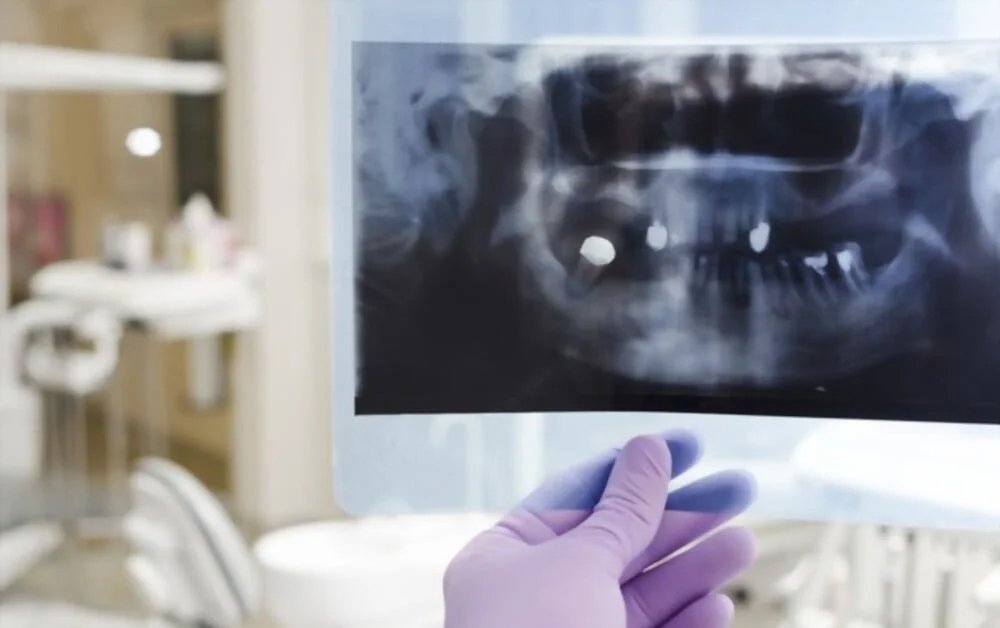
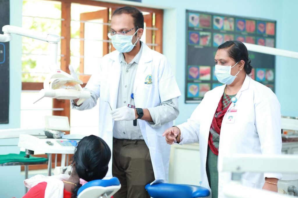
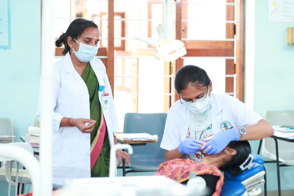
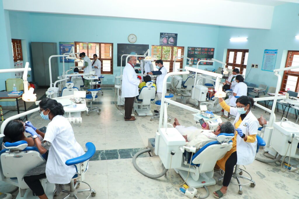
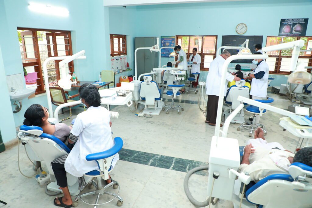
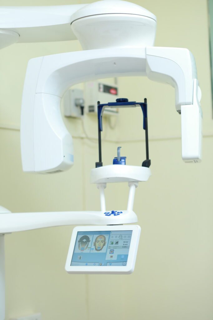
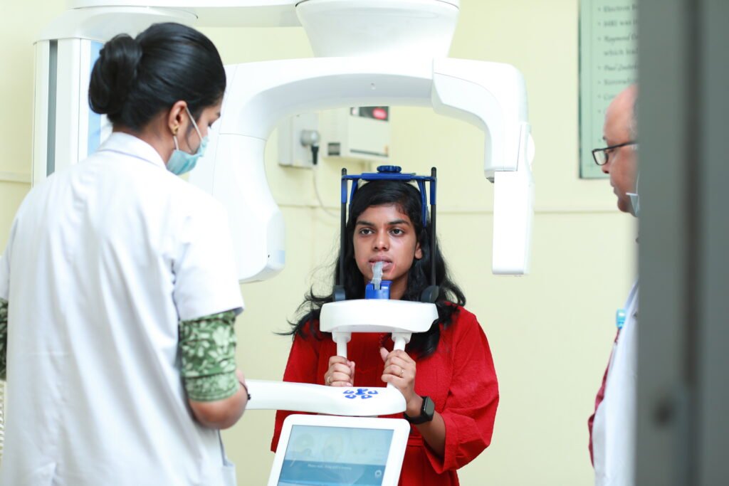
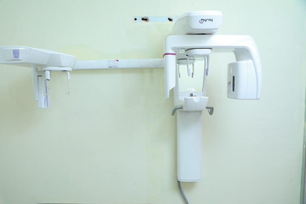
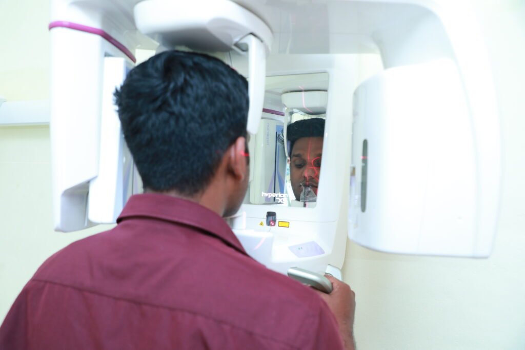
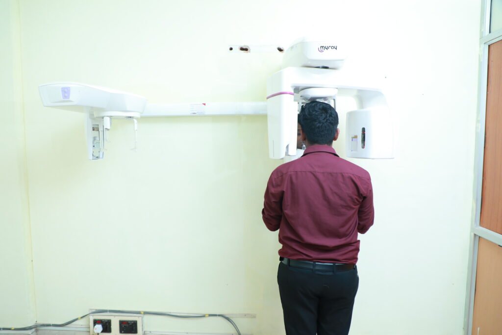
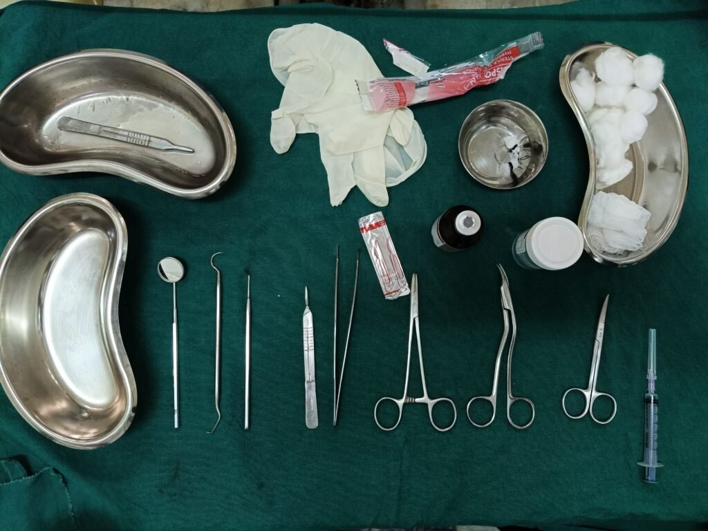
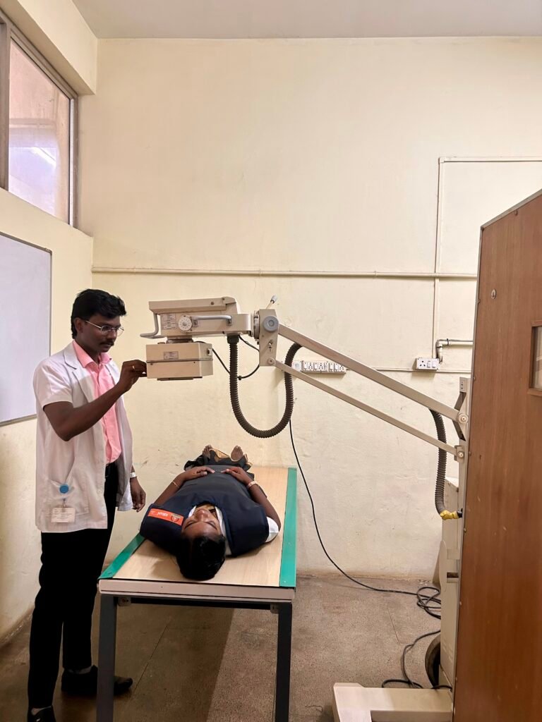
Our department is equipped with a dedicated infrastructure to accommodate advanced imaging equipment such as the CBCT machine and OPG machine.
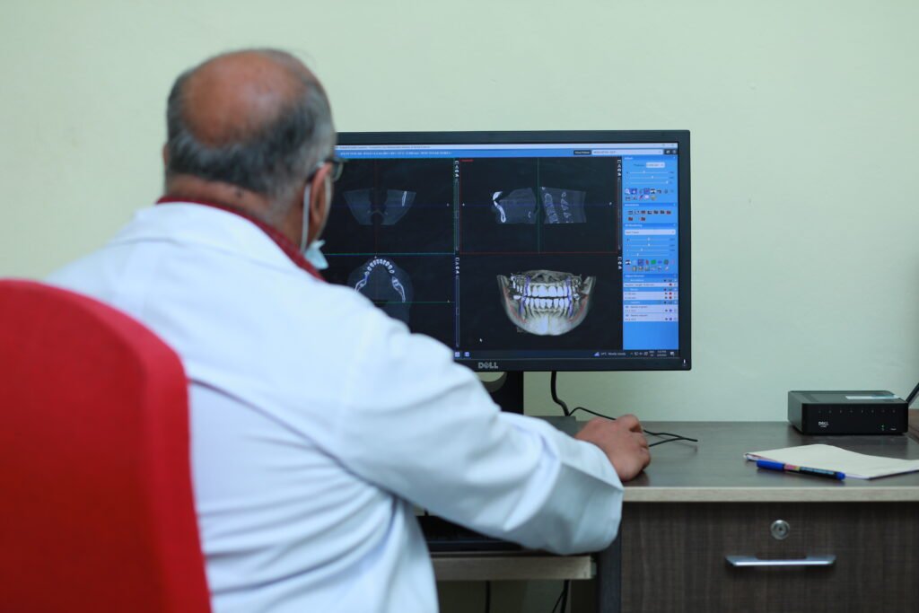
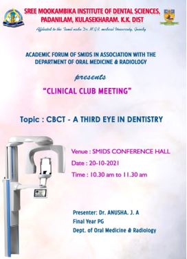
Cone-Beam Computed Tomography (CBCT) is an imaging modality that has shown a tremendous upward trend since its introduction in the late 1990s. The main advantage of CBCT technology is the ability to view and analyze in three dimensions (3D) the patient’s osseous oral and maxillofacial structures, overcoming the magnification and superimposition of structures found with 2D imaging modalities like panoramic imaging. The bone and calcified structures are visualized along with airway spaces. CBCT technology is being widely used in all fields of dentistry, including orthodontics, endodontics,oral surgery/pathology, periodontics, and implant treatment planning CBCT imaging provides high quality, accurate 3D representation of the osseous elements of the maxillofacial region.
Planmeca ProMax® (made in Finland)3D is a product family consisting of exceptional all-in-one imaging units. With up to five different imaging modalities these intelligent products can meet all modern maxillofacial imaging needs. Planmeca ProMax 3D units also offer a unique Planmeca Ultra Low Dose™ imaging protocol, enabling CBCT imaging with an even lower effective patient dose than standard 2D panoramic imaging.
2. TEACHING PROGRAMME
- Special lectures are conducted by eminent faculty of the specialty from other universities who are brought in to educate the students on specific topics.
- Problem based learning approach is adopted to give the students a “real life” experience of dealing with patients in clinical situations.
- Chair – side teaching methods are adopted to give the students a thorough clinical approach.
- Accessibility of staff for the students is important so that no student remains shy and introvert.
- A notice board is dedicated to the UGs and PGs which have to be managed by them in the way they deem appropriate, which is evaluated for content and quality by the staff and competitions with winner declaration being a regular practice.
- Feedback from the students and patients from time to time, helps us shape the future of the department.
- Student evaluation is conducted during theory and clinical postings by internal assessment (written, oral and practical). Internal assessment (Theory and Practical) exams are conducted for both undergraduates and post graduates.
- Regular intra department staff Meetings to ascertain our current academic progress and future plans. Systematic follow up on the programs envisaged and ensuring compliance.
3. PROCEDURE
- Diagnosis of oral cancer and precancerous lesions
- Identification of oral manifestation of systemic disorders
- Diagnosis and medical management of various oral diseases
- Diagnosis &non surgical management of temporomandibular disorders
- Medical management of orofacial pain
- Diagnosis & medical management of salivary gland disorders
- Management of medically compromised patients
- Minor biopsy procedures
4. SPECIALITY SERVICES
- Cone Beam Computed Tomography(CBCT) Machine-Planmeca Promax
- Digital extra oral radiographic machine – 100kVp
- Digital OPG & Ceph Machine
- PSP plates
- Intra oral X ray machine
- Intra-oral camera
- Facilities for Sialography
- Dormia basket for Lith retrieval
- Vital staining
- TMJ Clinic
- Fiberoptic Transillumination
- Tumor Board
5. CDEs, WORKSHOPS, EVENTS DONE IN THE DEPARTMENT
6. MERITS AND OTHER ACHIEVEMENTS
7. PUBLICATIONS
8.RESEARCH
Conclusion
The Department of Oral Medicine & Radiology at Sree Mookambika Institute of Dental Sciences is dedicated to fostering a comprehensive, patient-centered approach to oral healthcare. Through advanced diagnostic capabilities, innovative research, and exceptional educational programs, the department sets high standards in oral medicine, radiology, and neuromuscular dentistry, contributing to the growth and expertise of future dental practitioners.
Overview
Infrastructure
CBCT
OPG
Teaching Programme
Procedure
Speciality Services
CDE Program